Figure 4.
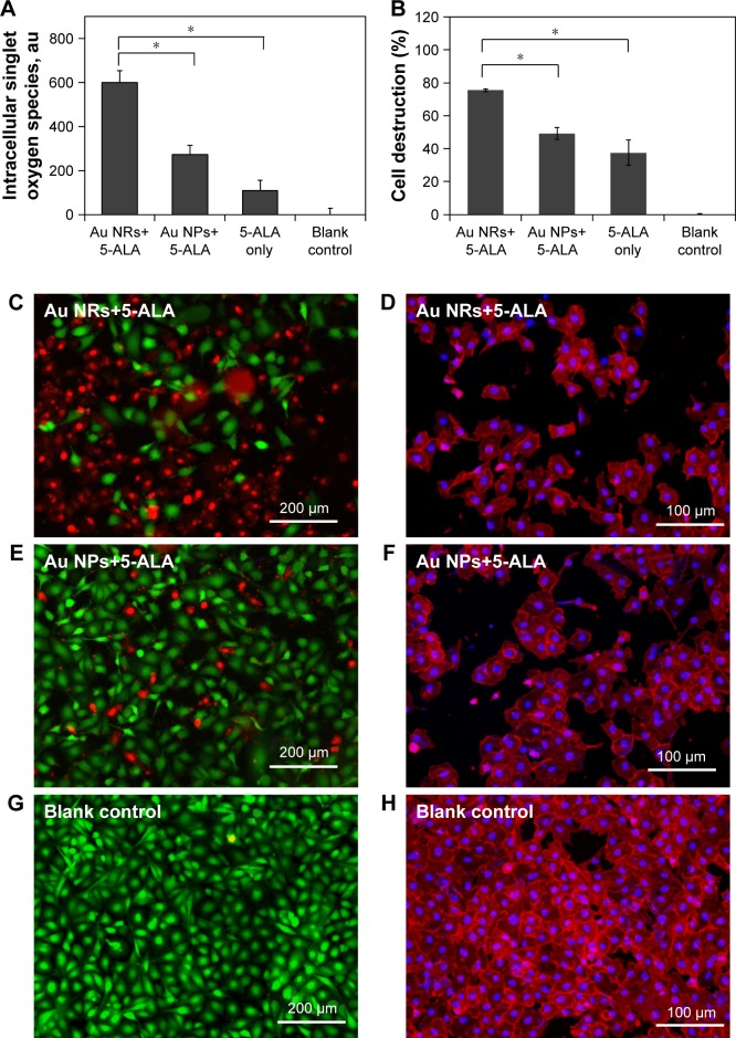
(A) Elevated generation of singlet oxygen in MDA-MB-231 cells upon various PDT treatments with broadband light irradiation for 1 min. (B) Cell destruction rate of MDA-MB-231 cells after various PDT treatments with broadband light irradiation for 1 min. Representative fluorescent images of MDA-MB-231 cells stained with a live/dead kit (left column, live cells stained green with calcein acetoxymethyl and dead cells stained red with ethidium homodimer-1), and sulforhodamine 101-conjugated phalloidin (right column, intracellular filamentous actin stained red and cell nuclei stained blue with DAPI) after various PDT treatments under broadband light irradiation for 1 min. (C) and (D) Au NRs+5-ALA, (E) and (F) Au NPs+5-ALA, (G) and (H) blank controls (no 5-ALA and Au nanostructures).
Notes: Prior to light irradiation, cells were treated with the combination of Au NRs and 5-ALA (Au NR+5-ALA), the combination of Au NPs and 5-ALA (Au NPs+5-ALA), and 5-ALA alone without Au nanostructures in serum-free medium. The final concentration of gold and 5-ALA as maintained at 40 µM and 1 mM for each group. Cells treated with serum-free medium without Au nanostructures or 5-ALA were considered as blank control. The data are representative of three separate experiments; *p<0.01.
Abbreviations: 5-ALA, 5-aminolevulinic acid; Au NP, gold nanoparticle; Au NR, gold nanoring; PDT, photodynamic therapy.

