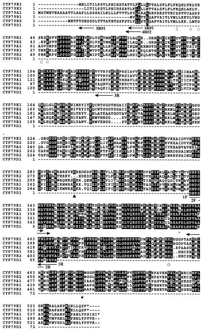
Figure 2.
Alignment of CYP79E1, CYP79E2, CYP79A1, CYP79B1, CYP79B2, and a PCR fragment of CYP79D1. Position of degenerate and gene-specific primers used to isolate the two CYP79s from T. maritima is indicated by arrows. Vector primers and the gene-specific primer for CYP79E1 5′-UTR are not shown. ●, Heme-binding Cys residue. ○, In the PERF region Phe is replaced by a His residue and Glu by a Asp residue. ×, In the conserved KETLR region, Leu is restored in CYP79Es. ▪, Met present in CYP79E2 but not in CYP79E1. □, Positively charged region in CYP79Es. ▴, Deletion in CYP79E2.

