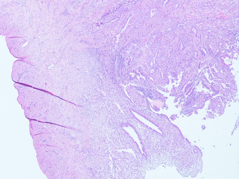
Figure 2.
Resection margin. Representative pathology slides showing endometrioid adenocarcinoma of the endometrium with polypoid pattern of growth and infiltration up to myometrial stroma. The resection margin is clearly assessable and appears free from disease. (Stained with hematoxylin/eosin, ×20 magnification.)

