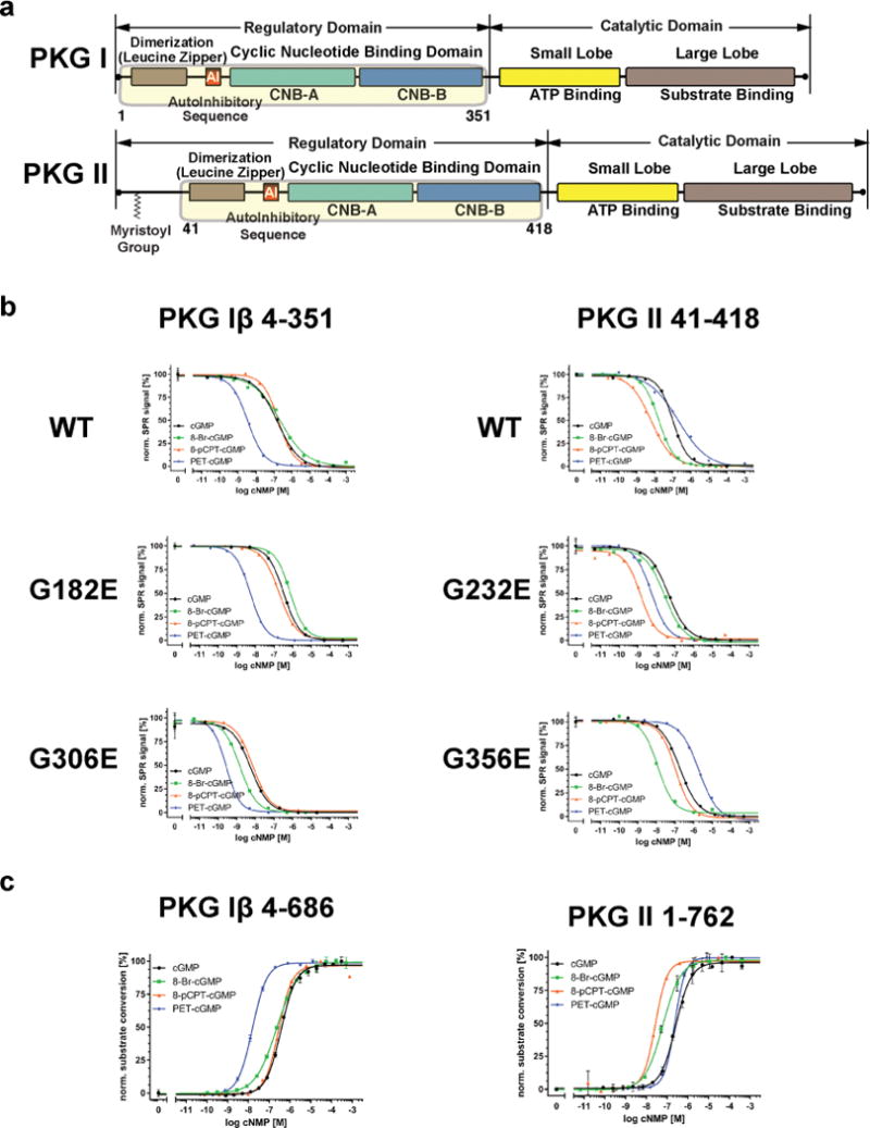
Figure 1.

Domain organization and SPR data. (a) The domain organizations of PKG I and PKG II are shown. The regulatory domains used for SPR are highlighted with corresponding residue numbers. (b) SPR measurements of the PKG Iβ and II R domains for cGMP and analogs. Competition SPR curves are shown for wild type and mutant proteins. (c) Activation of PKG Iβ and II full-length by cGMP analogs.
