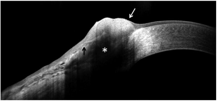
Figure 2.

An OSSN on HR-OCT with intermediate difficulty to interpret (some but not all classic findings). Note shadowing of inferior epithelial border (asterisk) and that the transition from normal to abnormal epithelium is not as distinct as in Figure 1. The transition from normal to lesion is rather distinct on the corneal side (white arrow), but less defined on the conjunctival side (black arrow).
