In Fig 2E, the wrong image appears in panel j and there are errors in the associated caption for panels g-i and j-l. Please see the corrected Fig 2 here.
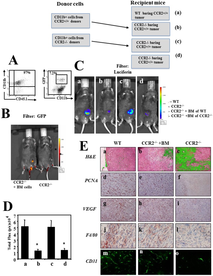
Fig 2. Bone marrow derived CD11b+CCR2+ cells are essential to support tumor development and angiogenesis.
(A) CD11b+ BMD cells from cx3cr1gfp CCR2+ CD45.1 mice were purified (left panel), analyzed fro the relative mummer of GFP+ cells (right panel) and transferred to CCR2−/− mice bearing CCR2+ tumor (B) shows imaging (IVIS) of a representative mouse as recorded using a GFP filter. (C) Imaging (IVIS) of the primary tumor on day 60, as recorded by the IVIS camera using–luciferin filter (recording luciferase activity of the cancer cells) as follows: CCR2+/+ C57BL/6 mice (WT) (a), CCR2−/− mice (b), CCR2−/− transplanted with BM of WT mice(c) and CCR2−/− transplanted with BM of CCR2−/−mice. All photos show a representative mouse per group (1 of 6 mice). (D) The computerized CCCD analysis of six mice per group. Results are shown as total flux (p/s ×104) ±SE. * Indicates p<0.001 (E) Histological, Immunohistochemical and immunofluorescence analyses of primary tumors from C57BL/6 WT mice, CCR2-/- (KO) mice, and CCR2-/- (KO) mice reconstituted with BM from WT mice. Panel a-c show H&E staining, d-f show anti -PCNA staining, g-i show anti VEGF, j-l show anti F4/80, m-o show anti CD31.
Reference
- 1.Izhak L, Wildbaum G, Jung S, Stein A, Shaked Y, Karin N (2012) Dissecting the Autocrine and Paracrine Roles of the CCR2-CCL2 Axis in Tumor Survival and Angiogenesis. PLoS ONE 7(1): e28305 https://doi.org/10.1371/journal.pone.0028305 [DOI] [PMC free article] [PubMed] [Google Scholar]


