FIGURE 2:
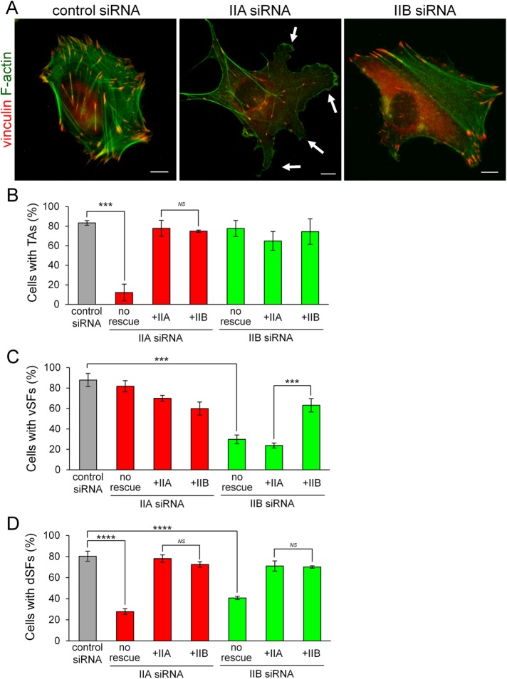
NMIIA and NMIIB are required for the formation of TAs and vSFs, respectively. (A) SV1 cells treated with the indicated siRNAs were fixed and stained with an anti-vinculin monoclonal antibody (red) and FITC-phalloidin (green). White arrows point to aberrant protrusions at the leading edge. Bar, 10 μm. (B–D) Rescue experiments of the defects in NMIIA-KD and NMIIB-KD cells on exogenous expression of each NMII isoform. Percentages of cells exhibiting TAs (B), vSFs (C), and dSFs connecting to TAs at right angles (D) on treatment with the indicated siRNAs. Note that NMHC-IIB rescued the defect in TA formation in NMIIA-KD cells, but NMHC-IIA failed to rescue the defect in vSF formation in NMIIB-KD cells. Data represent the mean ± SD from three independent experiments with n > 30 cells per experiment. ***p < 0.0005, ****p < 0.00005.

