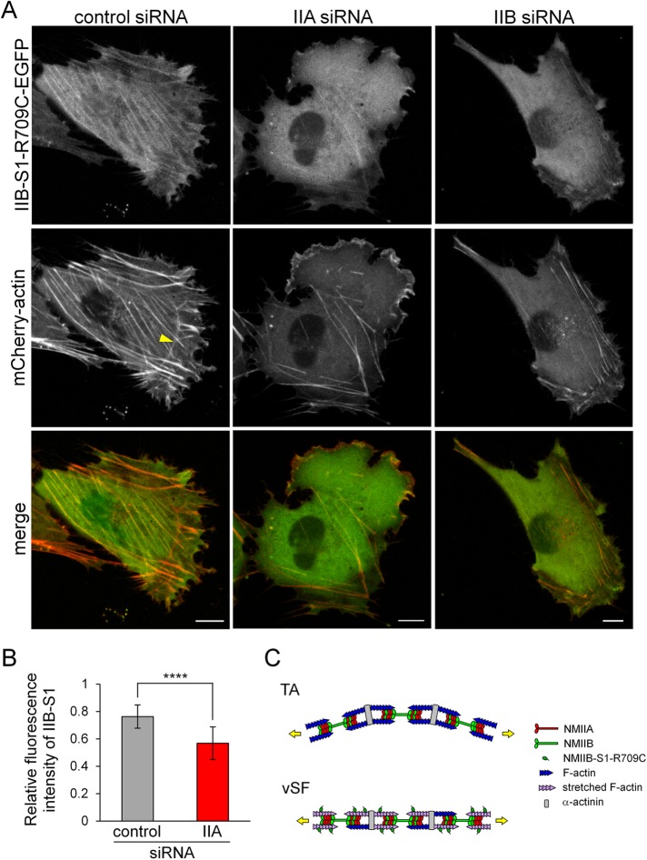
FIGURE 6:
Both NMIIA and NMIIB are required for the normal organization of vSFs. (A) SV1 cells treated with the indicated siRNAs were transfected with siRNA-insensitive NMIIB-S1-R709C-EGFP and mCherry-actin. Live cell images were captured using a confocal microscope. Bar, 10 μm. The yellow arrowhead points to a curved TA. Note that NMIIB-S1-R709C-EGFP did not bind to curved TAs. The fluorescence intensity of NMIIB-S1-R709C-EGFP relative to that of mCherry-actin in vSFs was lower in NMIIA-KD cells than in control cells. (B) The ratio of the fluorescence intensity of NMIIB-S1-R709C-EGFP to that of mCherry-actin in vSFs in control and NMIIA-KD cells. The relative fluorescence intensity of NMIIB-S1-R709C-EGFP in the SF area versus the control area was quantified and normalized by that of mCherry-actin using the RGB Measure plug-in of ImageJ software. Data represent the mean ± SD from n = 25 SFs from five cells. ****p < 0.00005. (C) Schematic model of the organization of SF subtypes. The key difference between TAs and vSFs is their contraction mode. Specifically, vSFs undergo isometric contraction (length remains constant), whereas TAs are shortened during centripetal flow. Therefore, if an equal load is applied to these structures, vSFs generate greater tension than TAs, resulting in the stretching of actin filaments, which is recognized by NMIIB-S1-R709C.

