FIGURE 3:
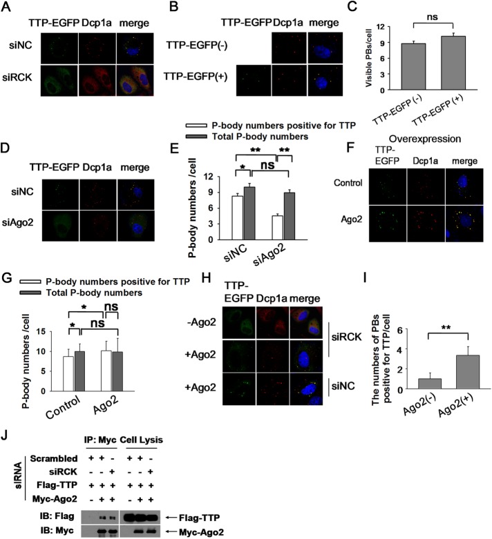
Ago2 is responsible for delivering TTP into PBs. (A) HeLa cells were transfected with siRCK to deplete the cytoplasm PBs. Scrambled siRNA (siNC) served as negative control. TTP-EGFP plasmid was also included in the transfection mixture. PBs were visualized with anti-Dcp1a antibody. (B) TTP overexpression did not increase the P-body numbers. HeLa cells were transfected with TTP-EGFP plasmid and the corresponding empty vector served as negative control. PBs were displayed by anti-Dcp1a antibody. (C) Graph showing the numbers of P-body as in B. Error bars represent standard error calculations obtained from averaging the PB number for 50 cells. ns, no significance. (D) Ago2 knockdown significantly reduced TTP localization in PBs. HeLa cells were treated with siAgo2 or siNC twice. TTP-EGFP plasmid was included in the second transfection mixture. PBs were shown by anti-Dcp1a staining. (E) Graph showing the P-body numbers that are positive for TTP, and the total P-body numbers per cell, in siNC and siAgo2 conditions. Error bars represent standard error calculations obtained from averaging the PB number for 50 cells. *p < 0.05; **p < 0.01; ns, no significance. (F) Ago2 overexpression significantly increased the localization of TTP within PBs. HeLa cells were transfected with TTP-EGFP with or without Myc-Ago2 cotransfection. PBs were visualized with anti-Dcp1a antibody. (G) Graph showing the P-body numbers that are positive for TTP and the total P-body numbers per cell. Error bars represent standard error calculations obtained from averaging the PB number for 50 cells. *p ≤ 0.05; ns, no significance. (H) Both TTP and Ago2 could organize microscopically visible granules formation. HeLa cells were treated with siRCK twice to deplete PBs. TTP-EGFP plasmid was included in the second transfection mixture with or without Myc-Ago2. PBs were displayed by anti-Dcp1a antibody. (I) Graph showing the P-body numbers that are positive for TTP per cell. Error bars represent standard error calculations obtained from averaging the PB number for 30 cells. **p ≤ 0.01. (J) Western blots of anti-Myc immunoprecipitates from RNase A–treated HEK 293T cells treated with siRCK or siNC (negative control) twice. Plasmids Flag-TTP and Myc-Ago2 were included in the second transfection mixture. Precipitates (left panels) and total extracts (input, right panels) were probed for the presence of coexpressed Flag-TTP and Myc-Ago2, as indicated.

