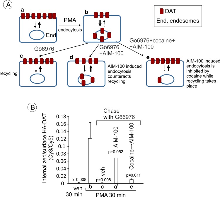
(
A) Schematics of the recycling assay and the interpretation of the results of such assay in (
B). Cells are incubated with DMSO (veh) (
a) or PMA at 37°C for 30 min to internalize a fraction of DAT (
b). At this point, most internalized DAT is in early endosomes, and thus is still capable of recycling (
Sorkina et al., 2003;
Sorkina et al., 2005). The cells were then chase-incubated in F12 medium with Gö6976 for 3 hr to block PKC activity and allow PKC-independent DAT trafficking (
c). The chase incubation of PMA-treated cells with Gö6976 at 37°C results in the gradual re-distribution of DAT from endosomes to the plasma membrane (recycling), where it is accumulated in ruffles, cell edges and filopodia. In the presence of AIM-100 during the chase incubation, DAT continues to internalize in an AIM-100-depenent manner (
d). If PMA- and Gö6976 treated cells are first preincubated with cocaine to prevent AIM-100-induced internalization of DAT and then chase-incubated with AIM-100, DAT translocated to the plasma membrane from endosomes, suggesting that AIM-100 does not inhibit the recycling of DAT from endosomes in this type of experiments. Widths of arrows roughly correspond to the predicted rate of internalization and recycling. Punctate arrows indicate a minimal rate. (
B) PAE/YFP-HA-DAT cells were incubated with HA11 antibodies for 45 min at 37°C. Cells were then incubated with vehicle (DMSO) or 1 µM PMA at 37°C for 30 min (corresponding to
A–b). The cells were then incubated in F12 medium with 5 µM Gö6976 for 30 min, followed by 2.5 hr at 37°C with 5 µM Gö6976 alone (
A–c), together with 20 µM AIM-100 (
A–d), or 10 µM cocaine and AIM-100 (30 min pre-incubation with cocaine and then AIM-100 for 2.5 hr) (
A–e). After fixation, cultures were stained with secondary anti-mouse antibodies conjugated with Cy5 (
surface HA-DAT), permeabilized with Triton X-100 and stained with secondary anti-mouse conjugated with Cy3 (
internalized HA-DAT). 3D images were acquired through 488 (YFP), 561 (Cy3) and 640 nm (Cy5) channels. Cy3/Cy5 ratios were calculated, and results are presented as mean values of the ratio (±SEM; n = 3-6). All P values are calculated in comparison with condition ‘b’ (cells treated with PMA for 30 min).


