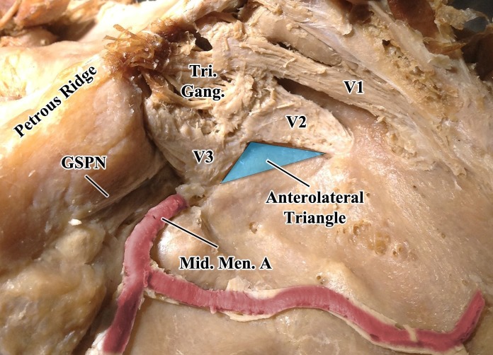
Figure 1. Superolateral view of the middle cranial fossa.
The dura mater has been removed to better visualize the underlying structures. The turquoise triangle represents the anterolateral cavernous sinus surgical triangle. The anterior border is defined along the posterior border of the maxillary division of the trigeminal nerve, from the intersection with the mandibular nerve to the posterolateral-most border of the foramen rotundum; the posterior border is defined along the anterior border of the mandibular division of the trigeminal nerve, from the intersection with the mandibular nerve to the anterolateral-most border of the foramen ovale; the lateral border is defined as a line connecting the posterolateral-most border of the foramen rotundum to the anterolateral-most border of the foramen ovale.
GSPN, greater superficial petrosal nerve; Mid. Men. A., middle meningeal artery; Tri. Gang., trigeminal ganglion; V1, ophthalmic division of the trigeminal nerve; V2, maxillary division of the trigeminal nerve; V3, mandibular division of the trigeminal nerve.

