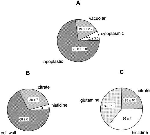
Figure 1.
Subcellular localization and speciation of Ni in leaves of hyper- and non-accumulator Thlaspi species as a percentage of total leaf Ni. Plants were exposed to 10 μm Ni in a hydroponic solution for 7 d. Leaf samples were collected and used for protoplast and vacuole isolation (A) or analyzed for Ni speciation using x-ray absorption (B). A, Subcellular localization of Ni in T. goesingense as determined by cell fractionation. For each independent replicate experiment, leaf Ni concentrations (as nmol g−1 fresh biomass) and protoplast Ni concentrations (as Ni per 106 structures; primary data as summarized in Table III) were normalized to chlorophyll contents. Ni in the protoplast fractions was calculated as a percentage of total leaf Ni, and the remainder of leaf Ni was concluded to be localized in the apoplast. Ni contents of the vacuolar fractions of the protoplasts were calculated based on the assumption that one vacuole was released per protoplast, and are expressed as a percentage of total leaf Ni. The remainder of the protoplast Ni was concluded to be localized in the cytoplasm. Values are averages of percentages calculated individually for three independent replicate experiments ± sd. B, Major Ni species present in T. goesingense as determined by x-ray absorption spectroscopy. Values represent the percentage of total leaf Ni associated with each ligand ± 95% confidence limit. Total leaf Ni was 501 nmol g−1 fresh biomass. C, Major Ni species present in T. arvense as determined by x-ray absorption spectroscopy. Values represent the percentage of total leaf Ni associated with each ligand ± 95% confidence limit. Total leaf Ni was 310 nmol g−1 fresh biomass. X-ray absorption data were collected from a single representative plant sample for each species and each x-ray spectrum used for the fits represents the mean of three independent scans, each being composed of data acquired from 13 independent detectors.

