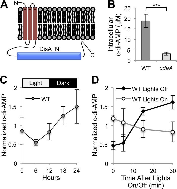
Fig 1. Presence, synthesis, and light-dependence of c-di-AMP in S. elongatus.
(A) Predicted CdaA protein topology. (B) Intracellular c-di-AMP measured by LC-MS for WT and cdaA transposon mutant (8S16-L9). Intracellular c-di-AMP concentrations were determined from raw quantities using cell volume (see Materials and Methods). The error bars represent standard error (SE) of five time points taken throughout a 24-h LDC in quadruplicate. ***P < 10−7 (Mann-Whitney-Wilcoxon Test). (C) C-di-AMP quantities in WT over one LDC. (D) C-di-AMP quantities upon the onset of darkness or light in WT. (C and D) C-di-AMP quantities are normalized by dividing by the average c-di-AMP concentration of the replicate. Error bars represent SE of four replicates.

