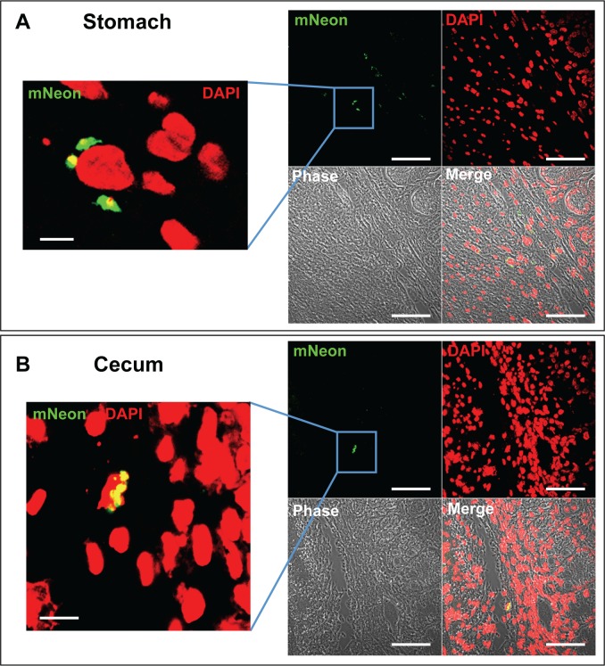
Fig 5. Location of parasites in the stomach and cecum during chronic stage infections.
A. Section of stomach tissue from a chronically infected mouse (day 117)—a single slice from a Z-stack through the infected cell (63x scan zoom 0.7). The left-hand panel is a magnified view of the boxed section (63x scan zoom 2.8). The bars represent 5 μm (left hand panel) and 50 μm (right hand panel). B. Section of cecum tissue from a chronically infected mouse (day 117), with a cluster of amastigotes surrounding the nucleus of an infected cell (left hand image, 63x scan zoom 2.5; right hand image, 63x scan zoom 0.7).

