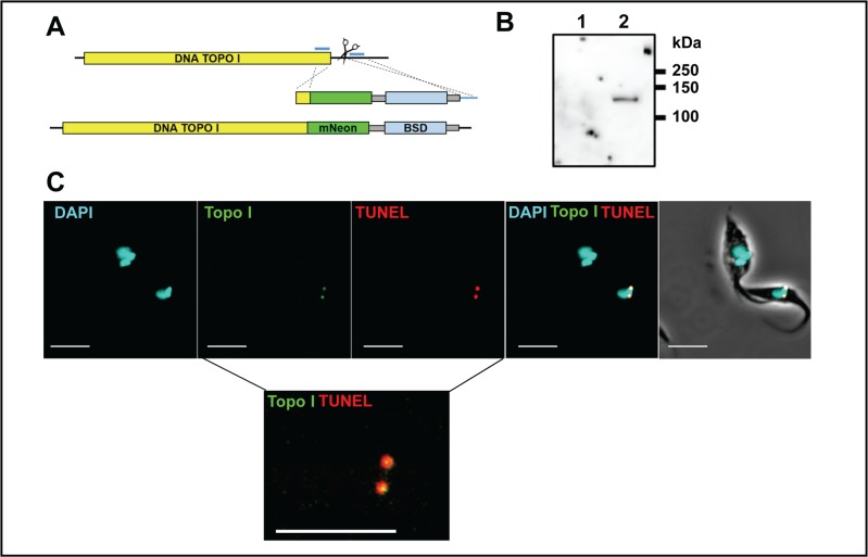
Fig 8. CRISPR/Cas9 mediated tagging and localisation of DNA topoisomerase 1A.
A. Map of the DNA topoisomerase 1A gene. The single sgRNA/Cas9 cleavage site is indicated by scissors. Blue lines indicate the positions of sequences corresponding to the 30 bp homologous arms in the repair fragment. The lower image shows the edited gene containing the 3’ fusion with mNeonGreen and the position of the selectable blasticidin (BSD) resistance gene. Grey boxes indicate sequences required for mRNA processing. B. Western blot showing the expression of the tagged DNA topoisomerase 1A. Lane 1, the CL-cas9 parental strain with an untagged locus; Lane 2, mNeonGreen tagged cell line showing a single band of the expected size (120 kDa). The blot was probed with anti-mNeonGreen (ChromoTek GmbH) C. Images showing that fluorescence is restricted to antipodal sites on the kinetoplast in parasites expressing mNeonGreen tagged DNA topoisomerase 1A. These sites coincide with regions of TUNEL positivity (Methods). Note that the upper parasite in the image, where the kinetoplasts have replicated and segregated, is negative for both mNeonGreen and TUNEL staining. The lower inset shows a magnified image where the mNeonGreen/TUNEL staining patterns are merged. Co-localisation appears as yellow. The bar indicates 5μm.

