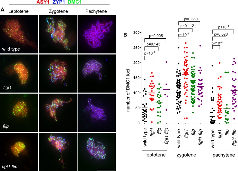
Fig 8. DMC1 foci in wild type figl1, flip and figl1 flip.
A. Triple immunolocalization of ASY1 (red), ZYP1 (blue) and DMC1 (green) on meiotic chromosome spreads. Merged pictures are shown. Partial and full ZYP1 polymerization defines the zygotene and pachytene stages, respectively. Scale bars 10μm. B. Quantification of DMC1 foci at leptotene, zygotene and pachytene in wild type, figl1, flip and figl1 flip. Each dot represents an individual cell and bars indicate the mean. P values are the results of Fisher's LSD tests.

