
Figure 1. Profiling retinal ganglion cell regeneration.
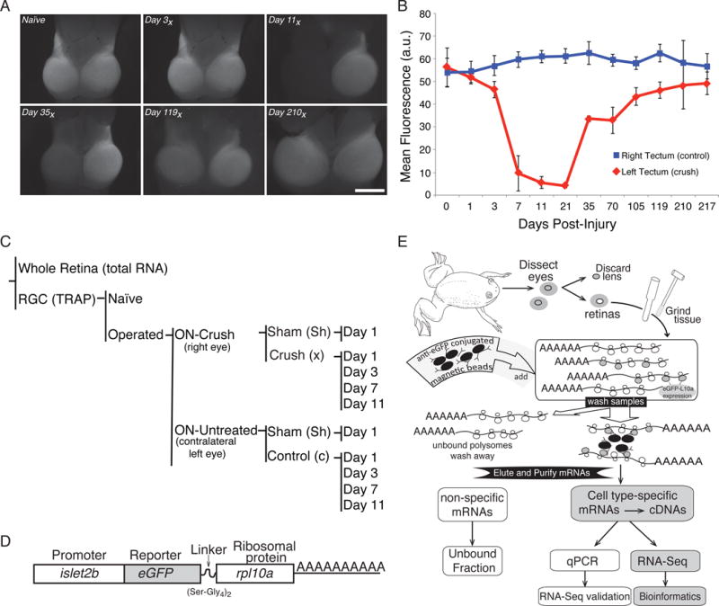
(A) The effects of axonal injury on retinal ganglion cells (RGCs) in the optic tectum can be visualized using frog lines expressing GFP under the control of an RGC-specific promoter (islet2b). An example time series shows the key transition point falls between 3 and 7 days post-injury, with full recovery occurring by 210 days (210x) post-injury. (B) Quantification of mean GFP fluorescence intensity in the tectum, as seen in panel (A). Data were averaged from at least 5 biological replicates per day and error bars represent the standard deviation from the mean. (C) In this study, gene expression in RGCs is directly compared between a right eye in which the optic nerve has undergone a surgical crush (Crush) to the untreated left eye of the same animal (Control) for various days after surgery (1, 3, 7, 11). Additional controls include a sham surgery (Sham), non-surgical animals (Naive), and RNA from whole retina (Total RNA). (D) To allow for tissue specific isolation of ribosome-associated mRNAs from RGCs, a transgenic line of Xenopus laevis is used that expresses an eGFP tagged variant of rpl10a under the control of an RGC-specific promoter (islet2b). (E) Following retina dissection, ribosome-associated RNAs in RGCs are purified using eGFP coated magnetic beads; subsequent poly(A) selection enriches for mRNA species. This mRNA fraction is then used for RT-qPCR validation and RNA-Seq library construction.