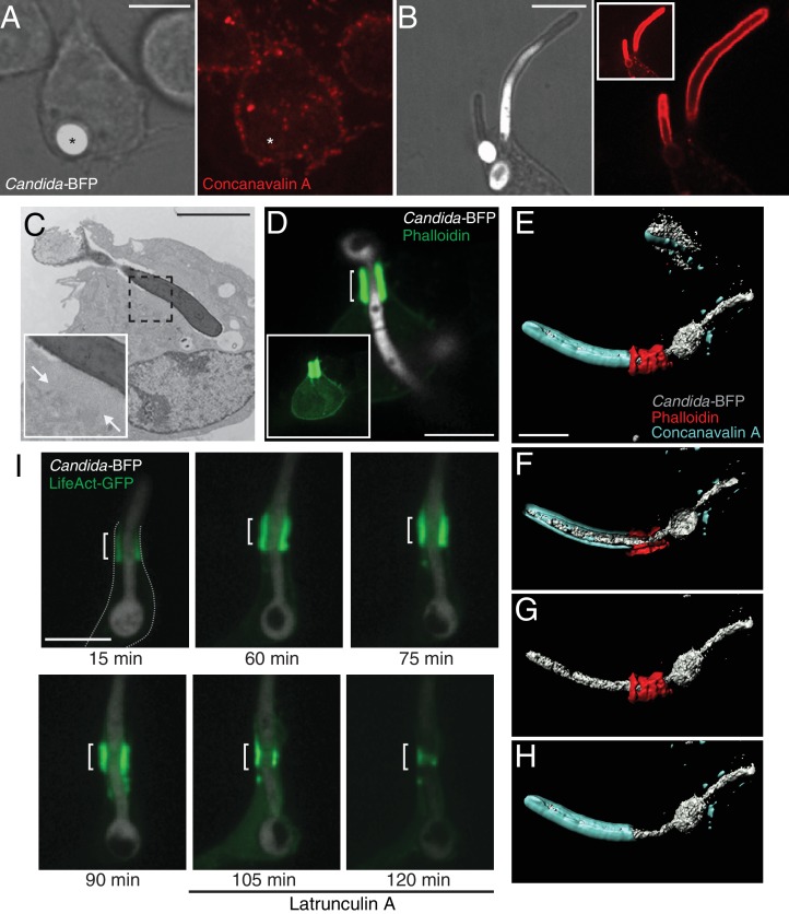
Figure 1. Partial phagocytosis of C. albicans hyphae is associated with formation of an actin cuff.
Phagocytosis of C. albicans yeast (A) or hypha (B) by RAW-Dectin1 cells. After incubation with Candida-BFP, RAW-Dectin1 cells were fixed and extracellular C. albicans stained using Alexa594-conjugated concanavalin A (red). The fluorescence of the BFP is shown in white here and elsewhere to reveal the location of the Candida-BFP. Inset in (B): overexposure of the concanavalin A signal to show less intense, staining of the macrophage membrane (as in A). Scale bars: 5 μm and 10 μm, respectively. (C) Transmission electron micrograph of a RAW-Dectin1 cell with a partially internalized C. albicans hypha. Area of organelle clearance corresponding to the cuff structure is indicated in inset by arrows. Scale bar: 5 μm. (D) F-actin enrichment at the neck of partial phagosome. RAW-Dectin1 cells were allowed to internalize C. albicans hyphae, fixed and stained with fluorescent phalloidin (green). Actin cuff indicated with a bracket. Inset: overexposure to show the less intense cellular actin. Scale bar: 10 μm. (E–H) 3D rendering of a C. albicans hypha partially internalized by a RAW-Dectin1 cell. After incubation with Candida-BFP (white), RAW-Dectin1 cells were fixed and extracellular portions of the hyphae stained using Alexa647-conjugated concanavalin A (blue). Actin was stained with fluorescent phalloidin (red). Scale bar: 5 μm. (F) 3D rendering sliced near the middle of the tubular phagosome, (G) same as E showing only the hypha (white) and actin (red), and (H) same as E showing only the hypha (white) and concanavalin A (blue). (I) Stability of the actin cuff assessed by live cell imaging. RAW-Dectin1 cells expressing LifeAct-GFP were allowed to internalize C. albicans hyphae and imaged at defined intervals. Where indicated (105 min) 1 µM latrunculin A was added and recording continued. Actin cuff location indicated by bracket. Scale bar: 10 μm. Images are representative of ≥30 fields from ≥3 separate experiments of each type. In this and subsequent figures the outline of the phagocyte (when not readily apparent) is indicated by a dotted grey line.


