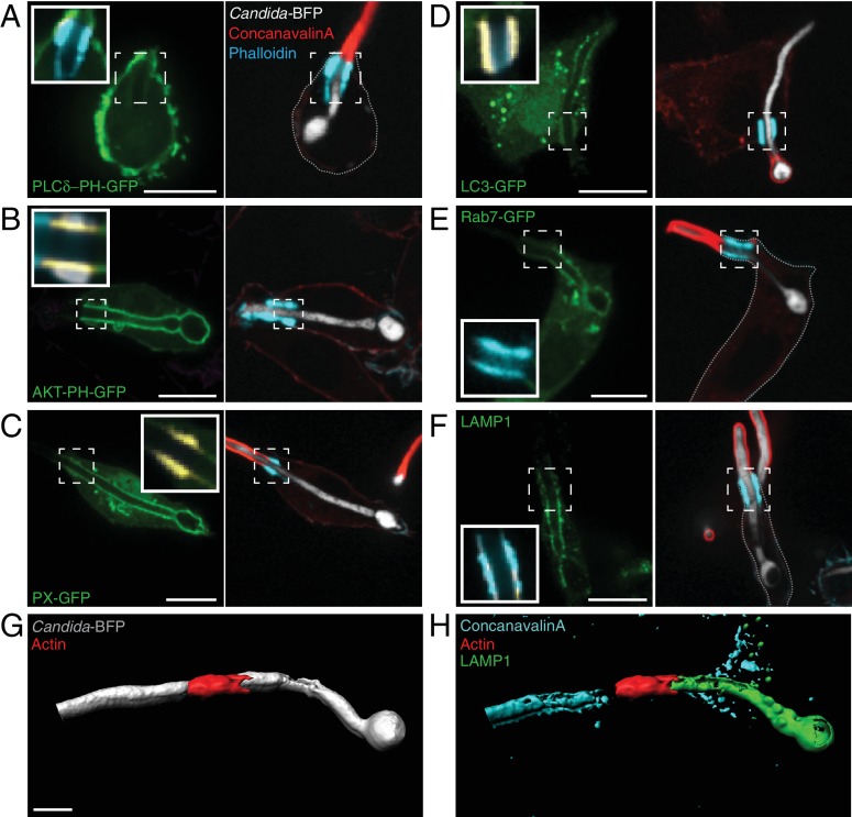
Figure 6. Distribution of phosphoinositides and endo-lysosomal markers.
After incubation with Candida-BFP hyphae, RAW-Dectin1 cells were fixed and extracellular C. albicans stained using Alexa594-conjugated concanavalin A (red). Actin was stained using fluorescent phalloidin (blue), and the location of the actin cuff is indicated by the dashed square. Visualization of: (A) PtdIns(4,5)P2 using PLCδ-PH-GFP; (B) PtdIns(3,4,5)P3/PtdIns(3,4)P2 using AKT-PH-GFP, inset: colocalization of actin cuff with AKT-PH, in yellow; (C) PtdIns(3)P using PX-GFP, inset: colocalization of actin cuff with PX, in yellow; (D) LC3-GFP, inset: colocalization of actin cuff with LC3, in yellow; (E) Rab7-GFP, inset: colocalization of actin cuff with Rab7, in yellow; (F) immunostained LAMP1 (green), inset: colocalization of actin cuff with LAMP1, in yellow. Scale bars: 10 μm. Images are representative of ≥30 fields from ≥3 separate experiments of each type. (G–H) 3D rendering of a RAW-Dectin1 cell with a partially internalized C. albicans hypha. After incubation with Candida-BFP, RAW-Dectin1 cells were fixed and extracellular portions of the hyphae were stained using concanavalin A (blue). (G) C. albicans (white) visualized with actin immunostaining (red). (H) Same 3D rendering as in (G), visualizing LAMP1 immunostaining (green), actin (red) and concanavalin A (blue). Scale bar: 5 μm.

