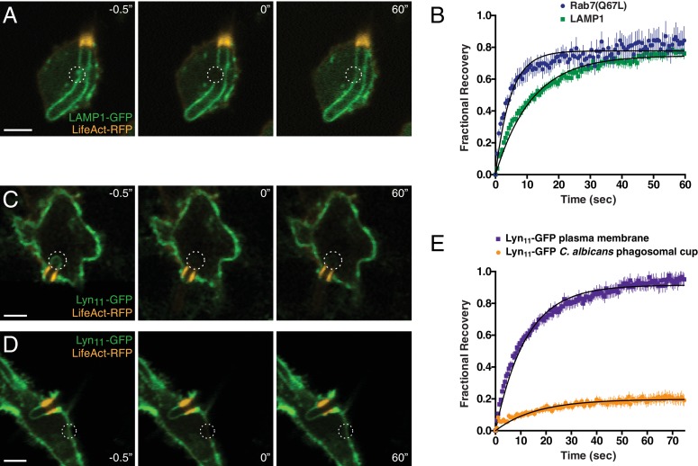
Figure 7. Formation of the actin cuff is associated with the establishment of a diffusional barrier.
RAW-Dectin1 cells were transfected with the indicated constructs, exposed to Candida-BFP, and used for FRAP determinations. F-actin was visualized with LifeAct-RFP (orange). (A) A region of interest (denoted by dotted circle) of LAMP1-GFP in the frustrated phagosome was selected (left panel, −0.5"), photobleached (middle panel, 0"), and allowed to recover for 60 s (right panel, 60"). Scale bar: 5 μm. Images in A, C and D are representative of ≥30 fields from ≥3 separate experiments of each type. (B) Quantitation of fractional recovery of fluorescence after photobleaching LAMP1 (green) or Rab7(Q67L) (blue). In both cases, data were normalized to fluorescence in unbleached regions of the C. albicans phagosomal cup. For either condition, four biological replicates, with a total of ≥30 cells, were quantified. (C–D) A region of interest in the frustrated phagosome (C) or in the plasma membrane (D) of cells expressing Lyn11-GFP was selected (left panel, −0.5"), photobleached (middle panel, 0"), and allowed to recover for 60 s (right panel, 60"). Scale bars: 5 μm. (E) Quantitation of fractional recovery of fluorescence of photobleached Lyn11-GFP in the plasma membrane (blue) or the frustrated C. albicans phagosomal cup (orange). In both cases, FRAP data was normalized to fluorescence in the plasma membrane. For either condition, three biological replicates, with a total of ≥35 cells, were quantified.


