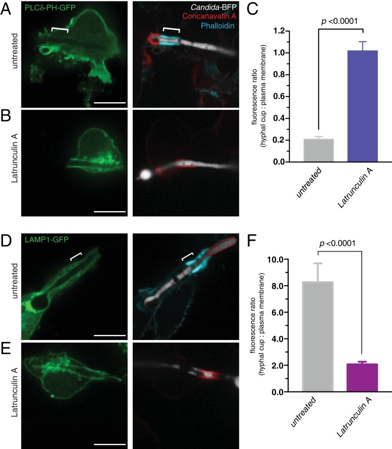
Figure 9. Actin depolymerization abolishes the diffusional barrier around C. albicans hyphae.
RAW-Dectin1 cells were transfected with the indicated constructs, exposed to Candida-BFP hyphae and incubated for 30 min in the presence (B and E) or absence (A and D) of latrunculin A. After treatment, cells were fixed and extracellular C. albicans stained using Alexa594-conjugated concanavalin A (red), and actin stained using fluorescent phalloidin (blue). (A and B) Cells transfected with PLCδ-PH-GFP. (C) Effect of latrunculin A on actin cuff-mediated segregation of PLCδ-PH-GFP to the plasma membrane, quantitated as the ratio of the fluorescence intensity of GFP in the phagocytic cup over the plasma membrane. (D and E) Cells transfected LAMP1-GFP. (F) Effect of latrunculin A on actin cuff-mediated segregation of LAMP1 to the frustrated phagocytic cup, quantitated as the ratio of the fluorescence intensity in the phagocytic cup over the plasma membrane. For A and D, location of the actin cuff is indicated with a bracket. Scale bars: 10 μm. Images are representative of ≥30 fields from ≥3 separate experiments of each type. For each condition in C and F, three independent experiments were quantified, with ≥10 fields counted per replicate. p value was calculated using unpaired, 2-tailed students t-test. Data are means ±SEM.

