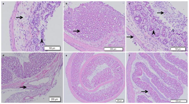
Fig. 6.
Deletion of Cldn7 in the large intestine induced by tamoxifen disrupted intestinal epithelial structure and induced hyperplasia. The arrow shows where the intestinal mucous membrane epithelial cells detached and necrosed (a). The glandular cavity was full of glandular epithelium cells, which were necrotic, and the glandular cavity almost disappeared. The mucosal epithelium cells and glandular epithelium cells were disorderly in arrangement and the goblet cells were smaller compared to the control group. The arrowhead indicates where local enteraden hyperplasia occurred (b). The arrows show where the mucous membrane epithelium cells were necrotic and detached and where the enteric cavity was mixed with a mucoid substance. The muscular layer was thinner (c). The arrow shows where connective tissue hyperplasia was phanic and broke through the bowel muscle layer to form hyperplasia, which included adipocytes, blood vessels, inflammatory cells, etc. (d). In the photograph, the normal intestinal structure of Cldn7 inducible conditional knockout mice was disordered (e). The intestinal epithelium structure of the solvent control group was intact and had no pathological changes (f).

