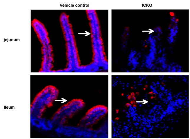
Fig. 9.
Immunofluorescent staining of the small intestine villi. The result indicates that obvious red fluorescence signals were arranged along the small intestine villi in the vehicle control group, while the ICKO mice showed weak and absent signals in jejunum and ileum, which means the structure and function of intestinal epithelial cells in Cldn7 ICKO mice was disrupted as shown by the arrowhead.

