Figure 5.
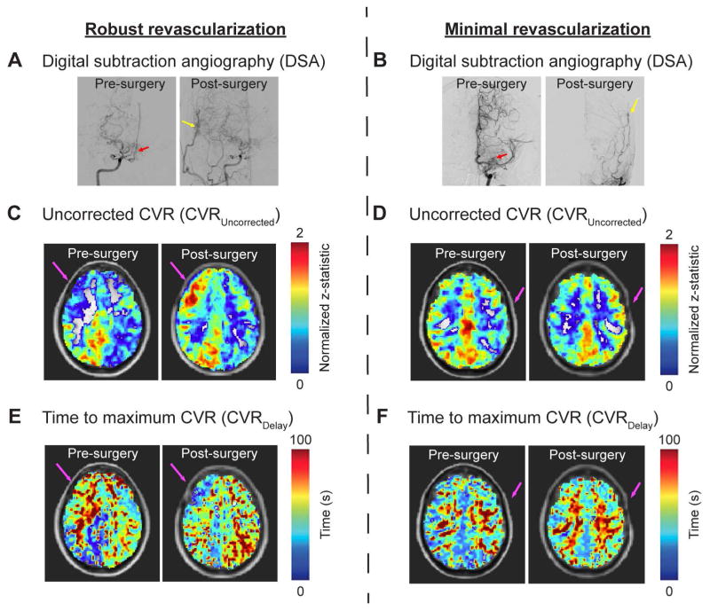
Blood oxygenation level-dependent (BOLD) MRI may be utilized to monitor treatment response in patients with moyamoya disease. (A, B) Representative examples of pre- and post-surgery digital subtraction angiography (DSA), (C, D) uncorrected CVR (CVRUncorrected), and (E, F) time-to-maximum CVR (CVRDelay) maps are shown for two patients: 1) a 28-year old Asian female with primary moyamoya disease and successful right-sided EDAS and 2) a 53-year old Caucasian female with secondary moyamoya disease and unsuccessful left-sided EDAS. Surgical success was determined based on whether 2/3 or more of the MCA territory was perfused following surgery as determined from DSA. For the first patient, success of the revascularization procedure is demonstrated by DSA, and corresponding increases in CVRUncorrected (C) and decreases in CVRDelay (E) can be seen. For the second patient, the revascularization procedure was unsuccessful based on the post-surgery DSA (B), and this is indicated by the lack of change seen in the CVRUncorrected (D) and CVRDelay (F) images. This example illustrates the potential of CVR imaging as a marker of treatment response to revascularization surgery in patients with moyamoya disease.

