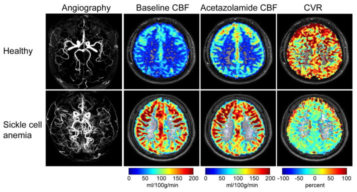
Figure 6.
Cerebrovascular reserve in sickle cell disease (SCD). (Above) Intracranial magnetic resonance angiography (MRA) and corresponding cerebral blood flow (CBF) maps at baseline and 10 minutes following intravenous acetazolamide (dose=16 mg/kg) infusion in a 24 year old healthy male without a history of cerebrovascular disease or hemoglobinopathy. CVR, defined as the fractional change in CBF in response to acetazolamide, is approximately 80% and largely symmetric throughout gray matter parenchyma. (Below) A 29 year old male with SCD and moyamoya syndrome. MRA shows hypervascularity and baseline CBF is elevated in response to anemia and reduced oxygen carrying capacity. Upon acetazolamide administration, the CBF response is blunted compared to the healthy control, translating to low-to-negligible CVR. These data are consistent with the SCD patient operating at or near cerebrovascular reserve capacity and such patients may be at elevated risk of future ischemic events. Images provided by Lena Vaclavu and additional information can be found in Vaclavu L et al. (Vaclavu et al., 2017).

