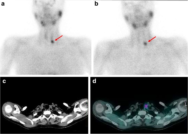
Fig. 1.
a, b 99mTc-Sestamibi dual-phase planar images show a focal tracer-avid lesion (arrows) below the left thyroid lobe on both the early (a) and delayed (b) images. c, d SPECT/CT images of the neck and mediastinum: transaxial CT (c) and fused (d) images show a small soft-tissue lesion with tracer avidity inferior to the left thyroid lobe

