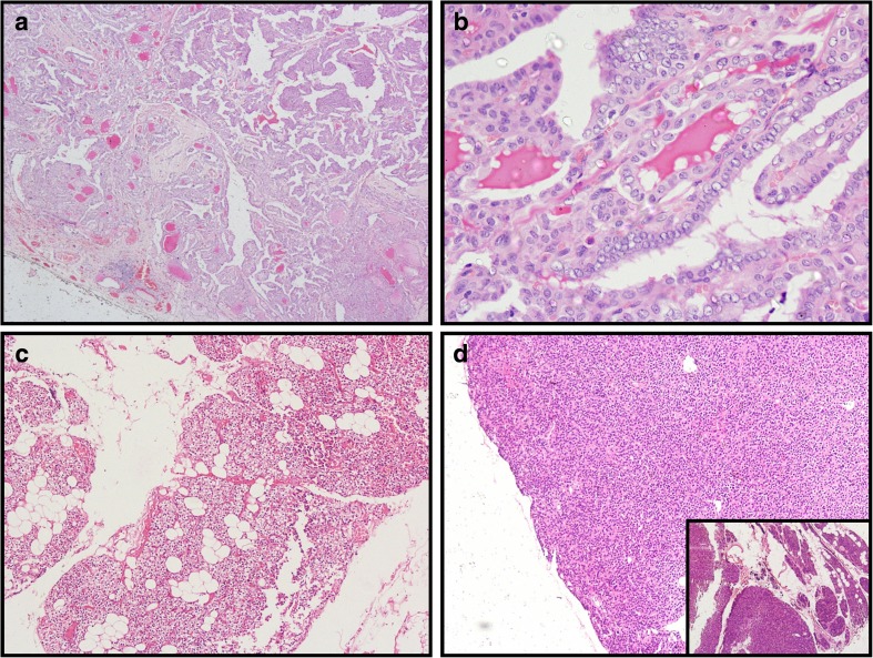
Fig. 3.
Histopathological examination of the surgical specimens. a, b Photomicrographs of H&E-stained sections show (a) the papillary configuration of the papillary thyroid carcinoma (×40) and (b) the nuclear features of the papillary thyroid carcinoma in the form of optical clearing, overcrowding and grooving (×400). c, d Photomicrographs of H&E-stained sections show (c) normal parathyroid gland (×100), and (d) normal parathyroid at the periphery of the adenoma (×40) and inset the adenoma

