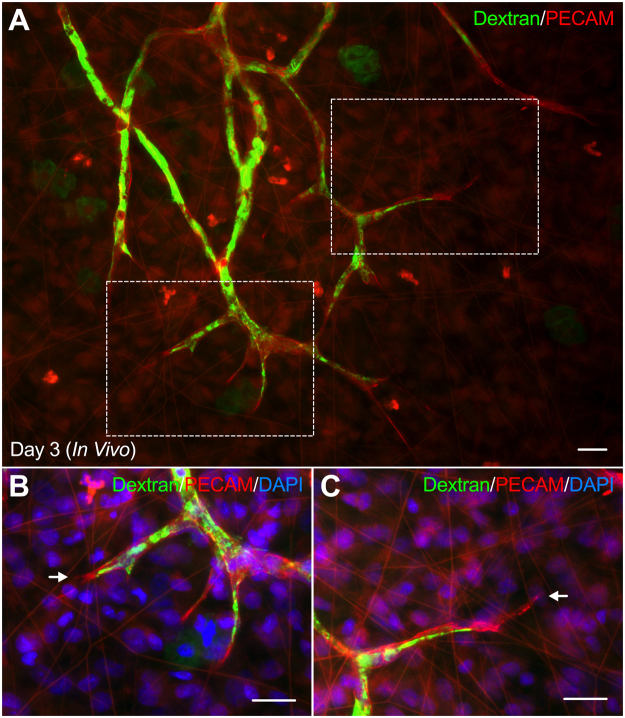
Figure 6.
Illustration of in vivo vascular lumens during angiogenesis. (A) Dextran perfusion of angiogenic in vivo microvascular networks identifies vascular lumens in capillary sprouts. (B,C) Higher magnifications of capillary sprouts in the tissue region, indicated by the above squares. Representative examples highlight the vascular lumen extending to the tip of a capillary sprout (B) as well as the vascular lumen forming behind the tip of the sprout (C). Arrows identify the distal edges of capillaries. Scale bar = 20 µm.

