Figure 7.
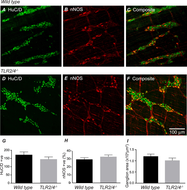
Immunohistochemical localisation of HuC/D (A,D) and neuronal NOS (B,E) in the myenteric plexus and circular muscle layer of distal colon from WT (A–C) and TLR2/4−/− (D–F). The composite images (C,F) illustrate the proportion of HuC/D positive neurons that also express nNOS. Scale bar in F applies to all panels. Summary graphs compare number of HuC/D positive neurons (G), proportion of nNOS positive neurons (H) and ganglion area (I) in distal colon of WT and TLR2/4−/−.

