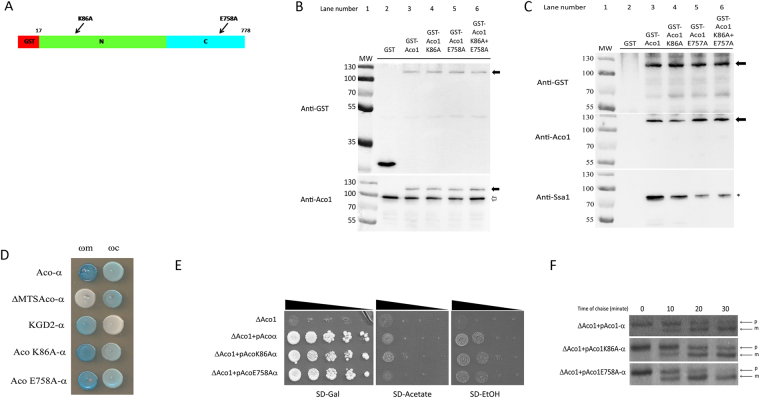
Figure 5.
Predicted Aco1 mutations reduce Aco1/Ssa1 binding. (A) Schematic representation of GST-Aco1 and mutations causing amino acid substitutions at the predicted Aco1/Ssa1 interface. GST tag, red; N terminal domains with the K86A mutation, green; C terminal domain with the E758A mutation, light blue. (B) Expression of GST-aconitase K86A and E758A mutant proteins. Total lysates of yeast cells expressing the indicated fusion proteins were subjected to Western blot analysis using either anti-GST (top panel) or anti-Aco1 (bottom panel). Arrows indicate plasmid encoded aconitase fusions, empty arrows indicates chromosomally encoded aconitase. (C) Pull down of Ssa1 by GST and GST-Aco1 mutants. The cell lysates above (B) expressing the indicated GST fusions were pulled down with glutathione sepharose beads and subjected to Western blot analysis using either anti-GST (top panel), anti-Aco1 (middle panel) or anti-Ssa1 (bottom panel) antisera. Arrows indicate the bands of GST-aconitase and its mutants, asterisk indicates bands of Ssa1. (D) Alpha complementation assay of aconitase-Ssa1 binding mutants. Yeast WT cells co-expressing cytosolic ω (ωc) or mitochondrial ω (ωm) together with the indicated Aco1-α mutants were grown on galactose medium containing X-gal. Blue colonies indicate alpha fragments that are associated with the ω fragments. Representative controls- pKgd2-α as a mitochondrial marker, pΔMTSAco1-α as a cytosolic marker. (E) Aconitase predicted Ssa1 binding mutants show different growth phenotype on acetate and ethanol medium plates. Aco1 KO yeast strains harboring the indicated plasmids were grown at the indicated dilutions on acetate, ethanol and galactose plates. (F) Mutations affecting the Aco1-Ssa1 interaction affect the efficiency of aconitase import. Aco1 KO yeast strains harboring the indicated plasmids were induced in galactose medium and pulse labeled with [35S] methionine–cysteine for 15 min, followed by a chase (addition of cold methionine–cysteine) for the times indicated. Total cell extracts were immunoprecipitated with aconitase antiserum and SDS-PAGE followed by autoradiography was performed. Precursor, p; Mature, m.

