FIGURE 1.

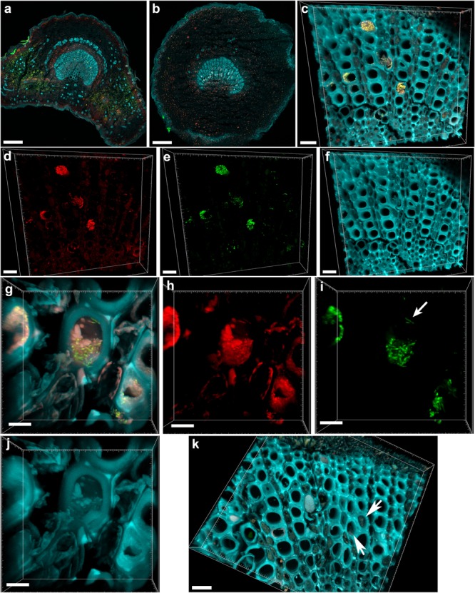
Confocal microscopy images showing the colonization of olive petioles by fluorescence in situ hybridization (FISH)-stained Xylella fastidiosa. (a,b) Maximum projections of confocal stacks showing basal leaf blade and petiole, respectively. (c) Magnification of the petiole vessels in the central part of section (b); yellow: X. fastidiosa, cyan: plant tissue autofluorescence; (c) is the overlap of (d–f). (d,h) Signal of the universal bacterial Cy3-labeled EUB338MIX FISH probe (red). (e,i) Signal of the X. fastidiosa-specific Cy5-labeled KO 210 FISH probe (green); arrow indicates a single X. fastidiosa cell with the typical rod-shaped morphology. (f,j) Autofluorescence of the plant tissue (cyan). (g) Particular of a X. fastidiosa-infected vessel showing bacteria (yellow) and extracellular matrix (pink); (g) is the overlap of (h–j). (k) FISH negative control of an infected petiole section, stained with Cy3- and Cy5-labeled nonsense probes NONEUB; arrows indicate the autofluorescence of occluded vessels (scale bars: 200 μm in a,b, 20 μm in c–f,k, 5 μm in g–j). These are representative images from several observations on five different infected trees.
