FIGURE 5.

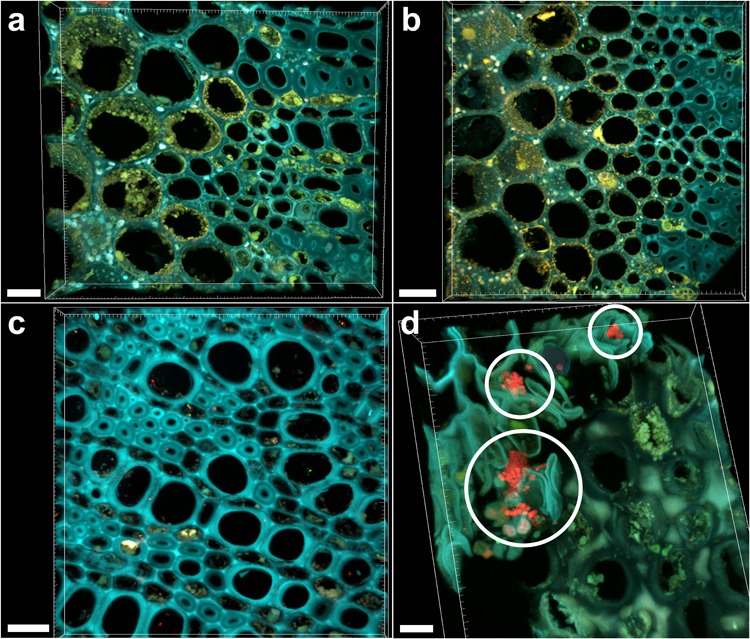
Confocal microscopy images showing FISH-stained branch sections. (a) Volume rendering showing the branch section of a Xylella fastidiosa-infected olive tree stained by FISH with the universal bacterial Cy3-labeled EUB338MIX and the X. fastidiosa-specific Cy5-labeled KO 210 FISH probes: all signals are autofluorescence. (b) Negative control of the branch section of a X. fastidiosa-infected olive tree stained by FISH with the Cy3- and Cy5-labeled nonsense probes NONEUB. (c) Branch section of an asymptomatic olive tree stained by FISH with the Cy3-labeled EUB338MIX and the Cy5-labeled KO 210 FISH probes: no occlusion of xylem vessels was observed. (d) Micro-colonies of other bacteria (not X. fastidiosa) detected in the branch sections of an infected olive tree, stained by the Cy3-labeled EUB338MIX probe (red; circles) (scale bars: 20 μm in a,c, 30 μm in b, 10 μm in d). These are representative images from several observations on five different infected trees.
