Figure 1.
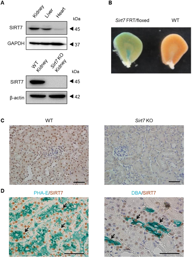
Expression of SIRT7 in the kidney. (A) SIRT7 expression in mouse kidney, liver, and heart was evaluated. GAPDH and β-actin were used as loading controls. SIRT7 protein expression was deleted in Sirt7 KO mouse kidney. (B) SIRT7 expression in the kidney of Sirt7 FRT/floxed mutant mouse was evaluated by β-galactosidase staining. (C) Representative photomicrographs (×400) of immunohistochemical staining for SIRT7 in kidney sections. Scale bar: 50 µm. (D) Representative photomicrographs (×400) of double immunostaining (lectin and SIRT7). PHA-E was used as a marker of proximal tubules and DBA was used as a marker of collecting tubules. The arrows indicate SIRT7-expressing nuclei. Scale bars: 50 µm.

