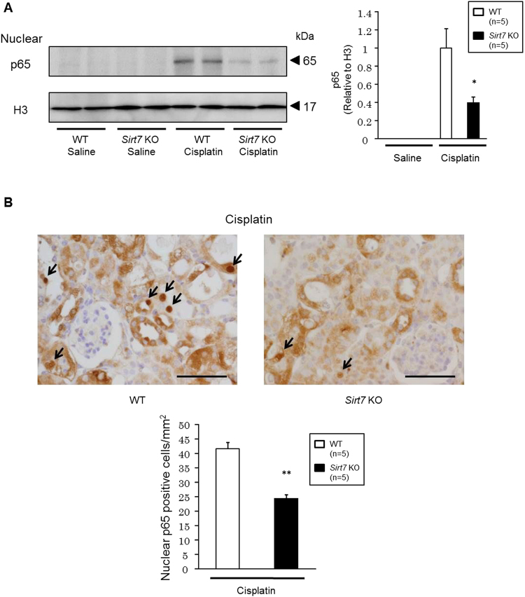
Figure 8.
Nuclear accumulation of p65 is suppressed in the kidney of cisplatin-treated Sirt7 KO mice. (A) Protein expression of p65 in the nuclear fraction of mouse kidney was evaluated by western blot analysis. H3 was used as a loading control (n = 5/group) (B) Nuclear p65 protein expression in tubular cells was evaluated by p65 immunostaining (n = 5/group). The arrows indicate p65-expressing nuclei. Scale bars: 50 µm. *p < 0.05, **p < 0.01. Data are expressed as the mean ± SEM.

