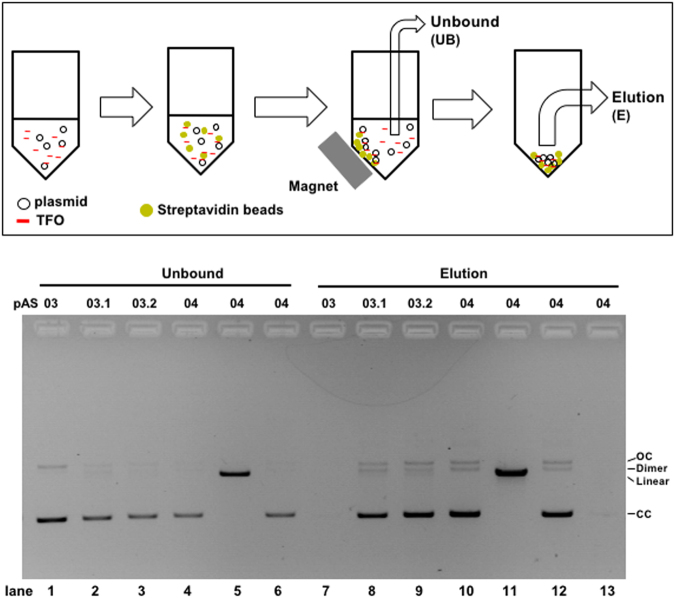
Figure 2.
Plasmid capture in vitro via TFO-mediated triplex formation: 600 ng (≈140 fmol) of each plasmid (pAS03, pAS03.1, pAS03.2, pAS04) are mixed with 8 pmol of TFO-1 for 24 hr, followed by mixing with streptavidin magnetic beads. The plasmid fraction not bound to beads (unbound: UB) is recovered using a magnetic stand (lanes 1–6). After the washing steps, plasmid bound to the beads is released either by boiling (lanes 7–11) or by competitive elution using a biotin-containing buffer (lane 12). Following competitive elution, the beads are re-suspended in a buffer and further eluted by boiling (lane 13). All plasmids are supercoiled circular except for lanes 5 and 11 where the plasmid is linearized. The recovered samples are analysed by agarose gel electrophoresis. Similar results were obtained in three independent experiments and a representative gel picture is shown here. In addition, TFO-3 was shown to perform similarly (data not shown). The loading amounts are 15 ng (lanes 1–6), 30 ng (lanes 7–12), and 60 ng (lane 13) plasmid equivalent. Estimation of plasmid recovery compared to input is: pAS03, <1% (lane 7); pAS03.1, ≈50% (lane 8); pAS03.2, ≈60% (lane 9); pAS04 (lanes 10–12), ≈70%, as quantified by NIH ImageJ software. CC: closed circular. OC: open circular. Dimer: dimerized plasmid.

