Fig. 2.
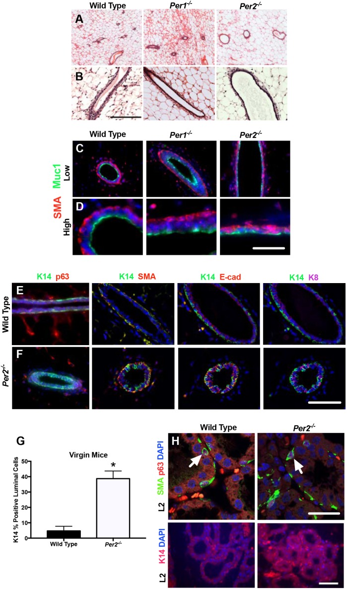
Altered expression of luminal and basal markers in Per2−/− mammary glands. (A,B) H&E staining of mammary gland sections from adult WT, Per1−/− and Per2−/− mice. (C-F) IF staining for myoepithelial and luminal cell markers, SMA and MUC1 (C,D), K14/p63, K14/SMA, K14/E-cad and K14/K8 (E,F), showing inappropriate localization of myoepithelial and luminal markers. (G) Quantification of K14-positive cells in WT and Per2−/− virgin mice (*P<0.05; error bars indicate s.e.m.). (H) IF staining of mammary gland sections in WT and Per2−/− mice at lactation day 2 (L2), showing colocalization (arrows) of SMA/p63 expression (upper panels) and normal K14 expression (lower panels). Per2−/−: n=4; WT: n=3. Scale bars: 50 μm.

