Fig. 4.

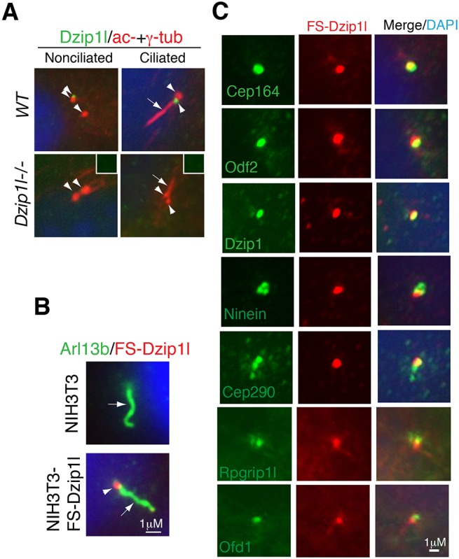
Dzip1l colocalizes partially with appendage markers of basal body and Rpgrip1l at the TZ. (A) Dzip1l localizes to the mother centriole in the cell. Wt and Dzip1l mutant pMEFs were immunostained for Dzip1l and acetylated α- and γ-tubulins (axoneme and centriolar markers, respectively). Dzip1l staining is positive in both ciliated and nonciliated wt mother centrioles, but not in mutant cells. Insets show negative Dzip1l staining in the centrioles indicated by arrowheads. Arrows point to cilia, and arrowheads indicate centrioles or Dzip1l staining. (B) The localization of overexpressed FS-Dzip1l recapitulates that of endogenous Dzip1l. NIH3T3 and NIH3T3-FS-Dzip1l stable cells were co-immunostained with Arl13b (labeling axoneme) and FLAG (for FS-Dzip1l) antibodies. (C) NIH3T3-FS-Dzip1l stable cells were co-immunostained for the indicated proteins and FS-Dzip1l (using FLAG antibody). Note the overlapping localization in yellow.
