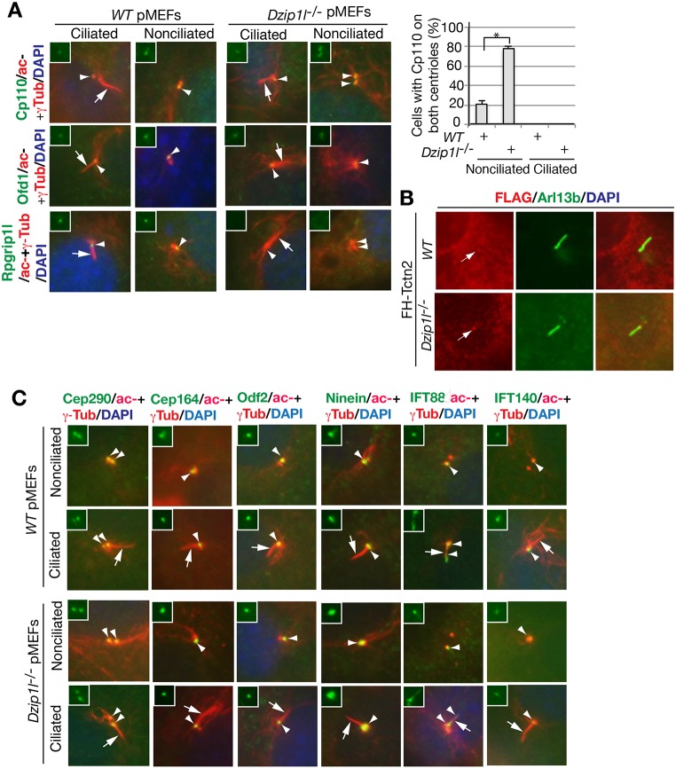
Fig. 8.
Dzip1l mutant cells fail to remove Cp110 from the mother centriole and to recruit Rpgrip1l to the mother centriole. (A) wt and Dzip1l mutant pMEFs were stained for the indicated proteins. The graph to the right shows the percentage of cells with Cp110 on both centrioles (n≥95). *P=0.0095, two-tailed Student's t-test. Note that Cp110 is not removed from the mother centriole and that Rpgrip1l is absent at the mother centriole. (B) Tctn2 localizes to the transition zone in Dzip1l mutant cells. Wt and Dzip1l mutant cells were transduced with murine retrovirus carrying FH-Tctn2 construct and then stained with FLAG (labeling FH-Tctn2) and Arl13b (a cilia marker) antibodies. Arrows point to FH-Tctn2 staining near the proximal end of cilia, presumably the TZ. FH, FLAG and HA double tags. (C) The localization of appendage proteins and recruitment of IFT proteins to mother centrioles are unaffected in Dzip1l mutant cells. Wt and Dzip1l mutant cells were stained for the indicated proteins and nuclei by DAPI. For both A and C, arrows point to cilia, and arrowheads indicate one or both centrioles. Insets show green staining for the indicated proteins at centrioles.

