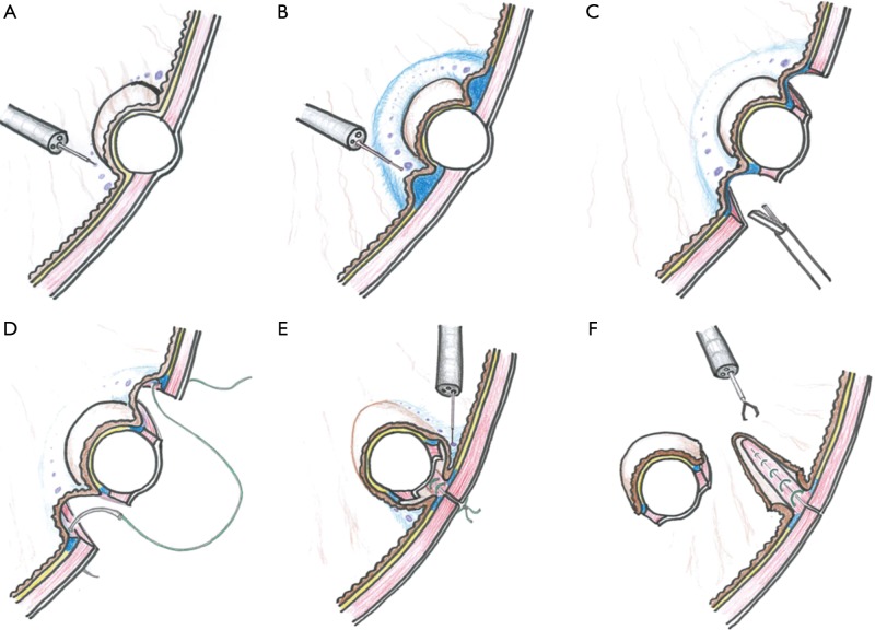
Figure 2.
Illustrations to explain the procedures. (A) Markings on the mucosa around the lesion; (B) injection into the submucosal layer; (C) circumferential seromuscular layer cutting; (D) seromuscular suturing; (E) incision of the muco-submucosal layer after inversion of the lesion; (F) loss of continuity between the lesion and gastric wall.

