Fig. 6.
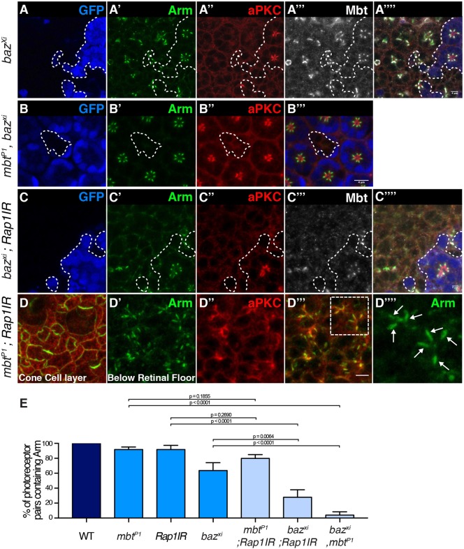
Rap1, Cno and Mbt synergize with Baz to promote AJ accumulation at the plasma membrane. (A–A‴′) bazxi106 mutant cells (lacking GFP, blue, the contour of which is denoted by the dashed line, A) and stained for Arm (A′), aPKC (A″) and Mbt (A‴). (B–B‴) mbtP1, bazxi106 double mutant cells (lacking GFP, blue, B) and stained for Arm (B′) and aPKC (B″). (C–C‴′) bazxi106, Rap1IR double mutant cells (lacking GFP, blue, C) and stained for Arm (C′), aPKC (C″) and Mbt (C‴). (D) Confocal section of the cone and pigment cells in an mbtP1; Rap1IR retina stained for Arm (green) and aPKC (red). (D′–D‴′) View of the delaminated photoreceptor proximal to D. (D′) Arm, (D″) aPKC, (D‴), merge (D″,D‴); a white-dashed rectangle highlights two ommatidia that are magnified in D‴′. The white arrows point to ZA domains between flanking photoreceptors. (E) Quantification of the percentage of pairs of photoreceptors sharing a lateral Arm domain in wild-type, mbtP1, Rap1IR, baz xi106, double mbtP1; Rap1IR, double baz xi106; Rap1IR and double baz xi106, mbtP1 cells. Results are mean±s.e.m. (n≥180 from 5 retinas). Scale bars: 4 μm.

