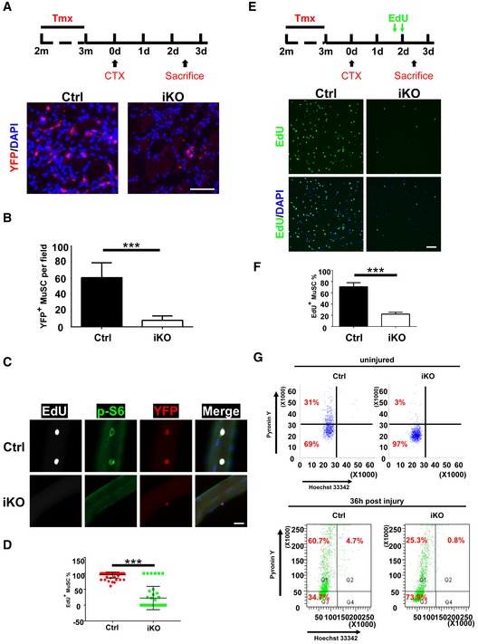
Adult littermates (
n = 3 in each group) of the Ctrl and iKO mice were injected with Tmx as described in Fig
1.
Top: the experimental scheme. 2.5 days post‐injury, TA muscle sections were immunostained for YFP (red). The nuclei were counterstained by DAPI (blue). m: month; d: day.
Quantification of the YFP+ MuSCs from (A) by counting ˜250 cells/mouse.
Freshly isolated single fibers (n > 20) were cultured with EdU for 48 h. Cells were then fixed and subjected to staining for EdU (white), p‐S6 (green), and YFP (red). Representative images are shown.
Quantification of the EdU+ MuSCs from (C) by counting > 20 fibers/mouse.
Top: the experimental scheme. Lower hindlimb muscles were injured by CTX followed by intraperitoneal injection of two doses of EdU at 40 and 48 h post‐injury. MuSCs from lower hindlimb muscles were sorted by FACS, fixed immediately and stained for EdU (green). The nuclei were counterstained by DAPI (blue). The representative images are shown.
Quantification of the EdU+ MuSCs from (E) by counting ˜500 MuSCs/mouse.
FACS‐isolated MuSCs from uninjured or injured muscles (36 h post‐injury) of the control and iKO mice were stained with Hoechst 33342 and Pyronin Y and analyzed by FACS.
Data information: Scale bars: 25 μm. In (B, D, F), the data are presented as mean ± s.d. ***
P < 0.001; unpaired Student's
t‐test.

