-
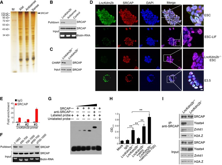
A
Biotin RNA pull‐downs were performed with nuclear extracts of mouse ESCs using full‐length lncKdm2b transcript (Sense), antisense, and Xist A repeats sequence control followed with mass spectrometry.
-
B, C
The interaction of SRCAP with
lncKdm2b was confirmed by immunoblotting (B) and CHIRP assay (C). Biotinylated probes were hybridized to
lncKdm2b, and chromatin complex was purified by magnetic streptavidin beads, followed by elution and immunoblotting. CHIRP probe sequences are listed in
Appendix Table S1.
-
D
Pluripotent ESCs and E3.5 blastocysts were probed with lncKdm2b by RNA‐FISH, followed by immunofluorescence staining for SRCAP. Green: lncKdm2b probe; red: SRCAP; nuclei were counterstained by DAPI. Scale bar, 10 μm. For normal ESC clone, n = 240; for LIF‐withdrawal ESC clone, n = 130; for lncKdm2b
−/− ESC clone, n = 105; for E3.5 embryos, n = 119.
-
E
Interaction of
lncKdm2b with SRCAP was verified by RIP assay. ESC lysates were incubated with anti‐SRCAP antibody, followed by RNA immunoprecipitation (RIP) assay. RNA was extracted and reversely transcribed.
LncKdm2b transcript was analyzed by real‐time qPCR. Fold changes were shown as means ± SD. Primers are listed in
Appendix Table S1.
-
F
Full‐length and truncated fragments of lncKdm2b were in vitro‐transcribed to biotin‐labeled RNA followed with RNA pull‐down and immunoblotting. d450–700 denotes the truncated fragment of lncKdm2b deleting nt 450–700. d700–1,000 denotes the truncated fragment of lncKdm2b deleting nt 700–1,000.
-
G
Nuclear extracts of ESCs and biotin‐labeled lncKdm2b (450–700 nt) probes were incubated for EMSA assays. Anti‐SRCAP antibody was preincubated with nuclear extracts that caused supershift.
-
H
ESC lysates were immunoprecipitated with anti‐SRCAP antibody, followed by detection of ATPase activities. Biotin‐labeled lncKdm2b and lncKdm2b (nt 450–700) fragments were generated by in vitro transcription by T7 RNA polymerase. Mouse IgG IP was used as a background control. Relative OD values were normalized to IgG background control and shown as fold changes as means ± SD. lnc, lncKdm2b; oe, overexpression. **P = 0.0034, **P = 0.0089, **P = 0.0023, **P = 0.0011 by unpaired Student's t‐test.
-
I
lncKdm2b
+/+ and lncKdm2b
−/− ESC cell lysates were immunoprecipitated with anti‐SRCAP antibody, followed by immunoblotting with the indicated antibodies.
Data information: All data are representative of five independent experiments.