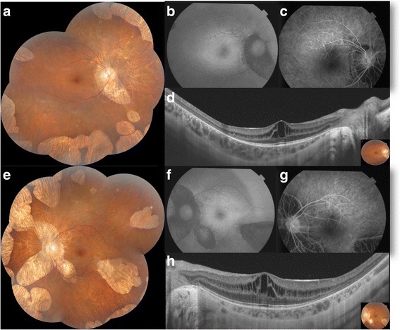
Fig. 1.

Case 1. Bilateral foveoschisis associated with gyrate atrophy. a,e. Composite colored fundus photography of both eyes showed multiple areas of well circumscribed chorioretinal atrophy in the midperipheral and peripheral zone. b,f. Fundus autofluorescence of both eyes revealed decreased autofluorescence in the areas of chorioretinal atrophy. c,g. Fluorescein angiography in the early phase (c) and in the late phase (g) didn’t reveal any leak in the macular area in both eyes. d,h. SS-OCT demonstrated a thickening of the macula with multiple hyporeflective spaces in the inner nuclear layer separated by vertical bridges suggesting foveoschisis in both eyes
