Fig. 2.

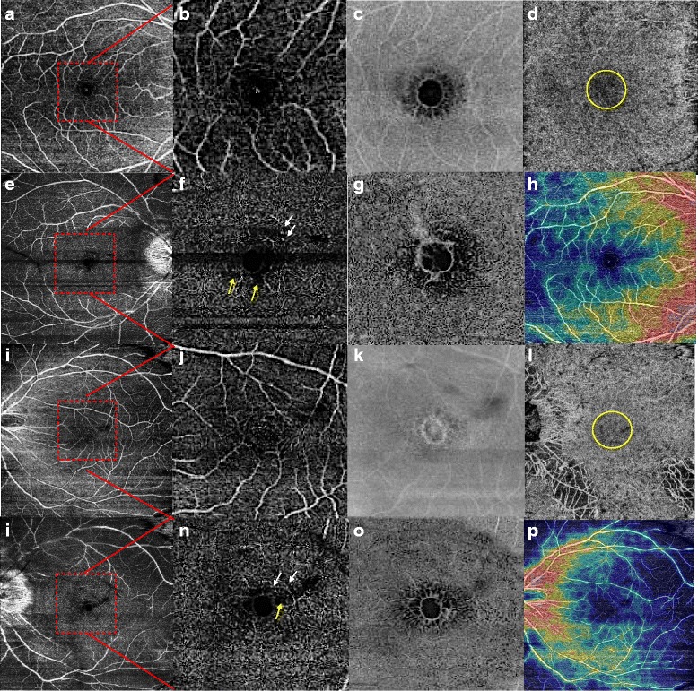
Case 1. Optical coherence tomography angiography and En-Face swept-source OCT in bilateral foveoschisis associated with gyrate atrophy. a,b,i,j. Superficial capillary plexus (9x9mm image) was normal in both eyes. The red square outlines the area of the 3x3mm magnified (b,j). e,f,m,n. Deep capillary plexus (e,m: 9x9mm, f,n: 3x3mm images) showed some perifoveal microvascular alterations similar to telangiectasias (white arrows) and petaloid non-reflective areas (yellow arrows). c,g,k,o. En-face images of superficial (c,k) and deep capillary plexus (g,o) of both eyes revealed a honeycomb pattern of the hyporeflective spaces better seen in the deep plexus. d,i. Choriocapillary layer in both eyes showed a central grey layer that could be due to a shadow effect (yellow circle). h,p. Density map of both eyes
