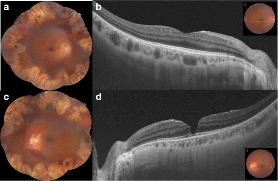
Fig. 3.

Case 2: Macular pseudohole associated with gyrate atrophy. a,c. Composite colored fundus photography of both eyes showed coalescent areas of peripheral chorioretinal atrophy sparing the posterior pole. b,d. SS-OCT was normal in the RE and revealed a macular pseudohole with a fine epiretinal membrane in the LE with a disorganised ellipsoid zone at the base of the pseudohole
