Fig. 1.

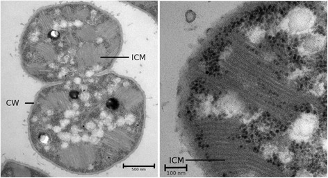
Transmission electron microscope picture of the strain Methylovulum psychrotolerans HV10-M2. Cell wall (CW) and intracytoplasmatic membrane (ICM) are labelled in the pictures. Scale bars represent 500 (left panel) and 100 (right panel) nm

Transmission electron microscope picture of the strain Methylovulum psychrotolerans HV10-M2. Cell wall (CW) and intracytoplasmatic membrane (ICM) are labelled in the pictures. Scale bars represent 500 (left panel) and 100 (right panel) nm