Fig. 1.

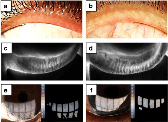
Representative photos and LipidView® II images of inactive (a, c, e) and active (b, d, f) thyroid eye diseases. a, b External eye photos of upper lid margins (OS). a) A 37-year-old female with inactive thyroid eye disease (CAS 1). Upper lid margin showed no pouting or capping of meibomian gland orifices. Only mild telangiectasia was observed. b) A 49-year-old female with active thyroid eye disease (CAS 3). Upper lid margin showed pouting and plugging of meibomian gland orifices. Telangiectasia was also present. c, d Infrared images of meibomian gland of the left lower lid. c) Grade 1 meibomian gland dropout (0−25%). d) Grade 2 meibomian gland dropout (25−50%). e, f Image of lipid layer (OS). e) Average lipid layer thickness: 79 nm f) Average lipid layer thickness: 100+ nm
