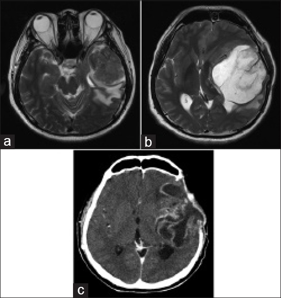
Figure 1.

(a) Magnetic resonance imaging scan of the brain showing the initial left temporal mass before surgery. (b) Magnetic resonance imaging of the brain showing tumor recurrence 5 months after initial surgery. (c) Computed tomography of the brain showing tumor recurrence 6 months after diagnosis and after two separate tumor resections
