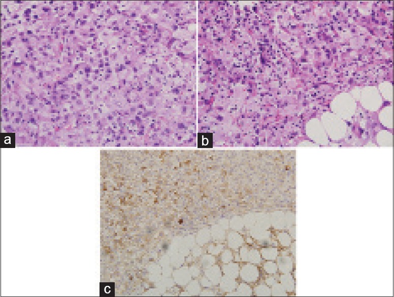
Figure 4.

(a) Histology of omental biopsy showing tumor composed of predominantly epithelioid cells with moderate amount of clear or eosinophilic cytoplasm and markedly pleomorphic, irregular, hyperchromatic nuclei with prominent nucleoli, consistent with anaplastic meningioma (H and E, ×400). (b) Cells with rhabdoid appearance characterized by eccentric nuclei displaced by rounded intracytoplasmic eosinophilic inclusions (H and E, ×400). (c) Positive immunostaining of the tumor cells with epithelial membrane antigen (×200)
