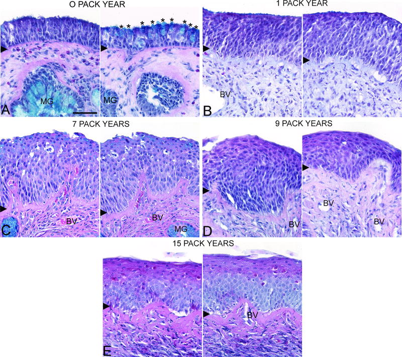
Figure 2.
Olfactory mucosal histopathology of CRS smokers with different pack year histories. Sections were stained with Alcian blue, hematoxylin, and eosin and two olfactory mucosal regions from the same biopsy section were imaged with a 40× objective. A. <1 pack year with normal olfactory epithelium and goblet cell hyperplasia, B. 1 pack year, C. 7 pack years, D. 9 pack years, and E. 15 pack years. Asterisks = goblet cells, MG = mucus gland, BV = blood vessel, and arrowhead = basement membrane. Scale bar = 50µm.

